
Annihilation (mutual destruction of matter and antimatter) has long been used in PET scanners. Now an interdisciplinary team from Poland has developed a lightweight and cheap new-generation tomography scanner - J-PET, which measures the rate of this annihilation in various tissues. This new biomarker makes it possible to create brain maps with completely new information, e.g. about glioma.
A team of physicists led by Professor Paweł Moskal from the Jagiellonian University published an article in the prestigious journal Science Advances. The paper discusses pioneering studies into the imaging of brain tumours conducted with the use of a new diagnostic parameter - the positronium lifetime. The team has developed the world's first glioma patient's brain image based on positronium.
Healthy brain tissue differs from that changed by cancer in terms of structure: for example, hydration, fat and oxygen content, intercellular spaces and the degree of 'packing' of molecules in cells. Scientists from Poland have shown that antimatter produced by radiopharmaceuticals - used in PET tomography - has a slightly different rate of annihilation in healthy tissue and in tissue changed by cancer. These are unimaginably small differences, on the order of tens of picoseconds (i.e. times hundreds of billions of times shorter than a second). However, they can be measured precisely enough to prepare a brain map on their basis.
This is completely new information that may be useful in diagnostics. Until now, PET only showed the place of radionuclide decay. The details of their disintegration process were not taken into account. The new generation of tomographs can provide additional information about changes in the brain during a single examination. 'It will be like a virtual biopsy', says Professor Ewa Stępień from the Jagiellonian University.
J-PET TOMOGRAPH: LIGHTER, CHEAPER, MORE SENSITIVE
This new generation of PET was developed some time ago by a team from Poland. 'We called our device J-PET - it is an abbreviation for Jagiellonian PET', says Professor Paweł Moskal. Professor Stępień adds that constructing a tomograph by individual research teams is something unprecedented on a global scale.
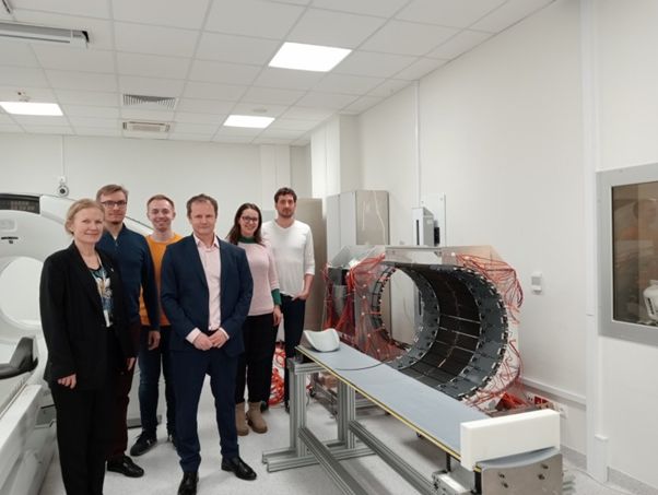
Standard tomographs are huge, heavy and expensive machines - they weigh several tons and are complicated to assemble. 'Our J-PET scanner weighs 60 kg. When we had to transport it for the duration of the study, we simply put it in the car, took it out, took measurements and packed it again', says Professor Moskal.
In standard PET, sensors recording the annihilation processes are based on heavy and expensive crystals. Meanwhile, the Polish device uses lightweight, cheap and more readily available plastics for this purpose. Thanks to this, the scanner is portable, easier to assemble and can be 10 times cheaper. An important novelty of this device is also that it captures more details regarding the annihilation process that are important from the diagnostic point of view.
ANNIHILATION IN THE BRAIN
First, let us explain what this annihilation used in PET is all about.
To prepare for the examination, the patient must receive radiopharmaceuticals intravenously. These drugs are constructed in such a way that they accumulate in the vicinity of the tumour. They contain isotopes that quickly undergo radioactive decay.
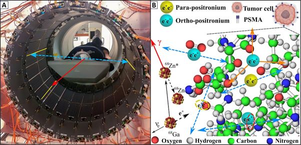
For example, when gallium-68 decays into zinc-68, it releases a positron, i.e. a particle of antimatter - it is like a mirror image of an electron, with a positive charge. If such a positron meets its antiparticle, an electron - annihilation occurs and two very strong photons are released.
These photons are so energetic that they pass through the human body, and the tomograph makes it possible to locate the place where they were created. On this basis, the tumour is located.
DANCE ME TO THE END OF LOVE
Professor Paweł Moskal has discovered that for diagnostic purposes, it is worth analysing the details of this annihilation process in even more detail.
This is because before a positron meets an electron and disappears forever, it often combines with it into an unstable structure - positronium. For a moment, positron and electron dancing around a common centre take on the characteristics of an atom - they are similar to hydrogen. Hydrogen also consists of a positive charge (proton in the nucleus) and a negative charge, but it is a stable structure that can last for billions of years. Meanwhile, positronium is an exotic atom that ceases to exist a moment after its appearance. The dance of this particle-antiparticle pair usually lasts 1-3 nanoseconds before the partners combine, and their mass (according to Einstein's famous formula) turns into energy.
It was precisely the time of this dance in various tissues that Professor Moskal measured using his J-PET. This was possible because J-PET is a multiphoton tomograph. It records not only the two photons produced during annihilation (like PET), but also an additional gamma quantum, which is produced a moment earlier, when the positron and electron begin their dance. It is the only scanner in the world that has this capability.
DANCE? NO PROBLEM
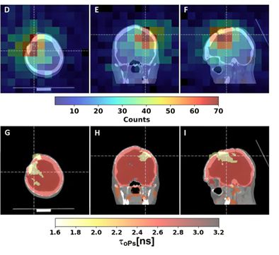
In the recently published study, it was determined that the lifetimes of a certain type of positronium in glioblastoma multiforme cells are shorter than in salivary glands and healthy brain tissue. In a brain tumour, it is an average of 1.7 nanoseconds, and in healthy brain tissue - 2.7 nanoseconds. The differences are unimaginably small, but still possible to notice.
'The lifetime of positronium tells us about the intramolecular spaces in which annihilation occurs', says Professor Stępień. The tighter this space is (as in the case of cancer cells), the faster the annihilation occurs.
DECAYING DRUGS
The researchers used previously clinically tested radiopharmaceuticals. They are also effective in imaging positronium, but the J-PET scanner does not use its full potential in this case. In the radiopharmaceuticals currently used, containing gallium-68, only 1 in a hundred electron-positron pairs signals that it starts dancing.
Professor Stępień points out that if a new pharmaceutical based on the decay of a scandium isotope (scandium itself is not new) produced from titanium were developed, it would be possible to record the length of the dance of 100% of positronium atoms, and the image would be more accurate. In addition, it would not be necessary to use compounds that typically accumulate around cancer, because positronium allows us to learn about the structure of the tissue, and not only - as in the case of PET - the place where the radioactive compound accumulates.
'We are already working on new pharmaceuticals with radionuclides for our PET. Now, if a hospital buys, for example, a gallium radionuclide generator for PET examinations, it has to replace it every nine months. If a radionuclide were prepared based on titanium and scandium, the hospital could buy a can of radionuclide, and its durability would be 60 years', says Professor Stępień.
Portable and cheap J-PET combined with highly durable radionuclides would make tomography much more accessible to everyone and could also reach poorer countries', she adds.
For now, the tomograph is intended for head examinations. 'The next step that we are planning is to build a larger J-PET, for whole-body examinations. The advantage will be that it will be possible to take an image in every tissue at once, and even prepare videos showing how the pharmaceutical is metabolised in different organs of the body. This is completely new information. Time and further research will show what to do with this information', says Professor Moskal.
The publication in Science Advances is one of many publications in prestigious scientific journals concerning J-PET. Professor Moskal has several dozen patents connected to the solution.
PAP - Science in Poland, Ludwika Tomala
lt/ zan/ kap/
tr. RL