Tendons of dinosaurs underwent the ossification process similarly to birds, shows research by Polish palaeontologists, published in the Zoological Journal of the Linnean Society.
Preservation of soft parts such as collagen fibres, blood vessels and cells is rare in the fossils of extinct vertebrates. Polish palaeontologists were the first to identify soft elements in dinosaur tendons.
The team also found that the soft tissues ossified in dinosaur tendons. This phenomenon is observed today in birds, when the soft tissues of the tendons mineralise during the life of an individual.
Team leader Dr. Dawid Surmik from the Institute of Earth Sciences of the University of Silesia in Katowice said: “The results of our research prove that dinosaurs and modern birds share the pattern of the tendon mineralisation process and the process of cell and tissue differentiation.”

Tendons connect muscles to bones. In most vertebrates, they are pliable and flexible, but in dinosaurs, including birds, some tendons underwent a process of ossification.
Dr. Surmik explains that in dinosaurs, as in birds, ossification of tendons led to the formation of bone-like structures, such as secondary osteons and bone-like cells (responsible for the exchange of nutrients in the bone).
He said: “This happened despite the fact that tendons before ossification are made of fibrous connective tissue, not cartilage, as is the case with most bones before ossification. It was previously suggested that only bone-like structures were present in ossified tendons, but this view can now be rejected.”
This means that tendons, although they are usually made of flexible and durable connective tissue, can ossify, transform into bone tissue. Such ossified tendons are found in modern birds, e.g. turkeys, in those parts of the musculoskeletal system that are exposed to heavy loads.
Dr Surmik said: “Tendon ossification may have occurred early in the evolution of certain groups of archosaurs (ruling reptiles), including pterosaurs, dinosaurs and modern birds, as an adaptation for more efficient locomotion or weight distribution.”
He added that the biggest challenge during the research project was the limited number of samples and the preparation of very small fossilised structures.
The analyses were destructive, associated with irreversible damage to the tested material. Surmik said: “Therefore, the minimum number of samples was used, and the sequence of analyses was planned very meticulously to use as little valuable material as possible, achieving the most objective results.”
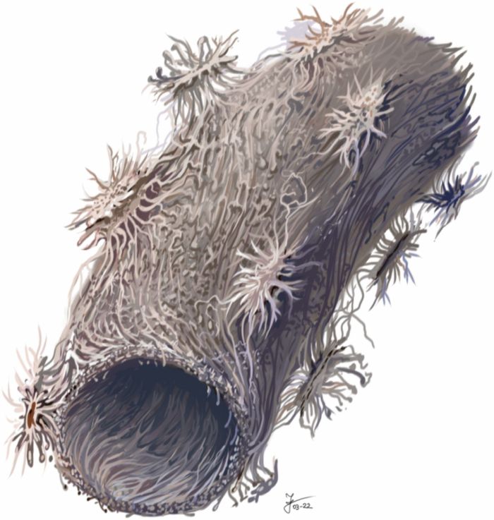
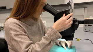
Samples of fossilized tendons were dissolved in a special chemical solution to remove phosphate, which is the main mineral component of bone. As a result of the dissolution of the bones, numerous very small, only a tenth of a millimetre in size, petrified fragments of blood vessels were obtained. They were accompanied by cells attached to them, with an average size below 0.01 mm. Precise studies of such tiny structures required the use of microscopes, including a scanning electron microscope and an atomic force microscope.
Dr. Surmik said: “Fossilized soft parts (vessels, cells, collagen fibres) in fossil vertebrate bones from the Mesozoic (252-66 million years ago) have been described since the 1960s, and the very fact of the preservation of structures resembling bone cells in cross-sections of fossilized bones of the examined under the microscope was noted by researchers in the 19th century.
“Recently, thanks mainly to the spectacularly preserved vertebrate fossils from China, cells have been identified in fossilized cartilage tissues. Never before, however, had fossilized tissues been searched for in the ossified tendons of non-avian dinosaurs. We searched and found them, and they were preserved in perfect condition!.”
Find out more in the source publication. (PAP)
krx/ agt/ kap/
tr. RL


