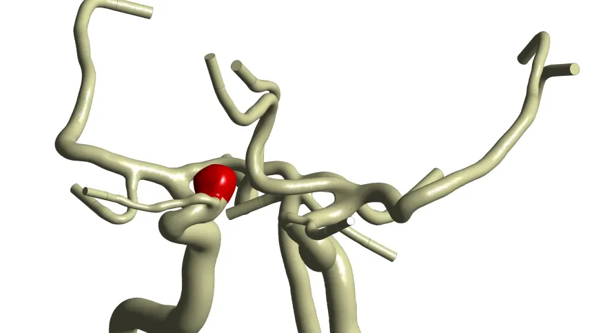
Aneurysms of various shapes and sizes, previously imaged in patients, will be virtually 'operated on' in many possible ways. This will lead to the creation of software that will help neurosurgeons secure them against breaking and growing.
'The attending physician will be able to choose the optimal variant of aneurysm treatment based on the anatomy of the patient's blood vessels. The tool will obtain results thanks to a properly trained neural network. The network learns on tens of thousands of data sets from advanced numerical blood flow simulations,’ says Dr. Zbigniew Tyfa from the Institute of Turbomachinery at the Lodz University of Technology, head of the team set up under the Polish National Centre for Research and Development LIDER programme.
According to the scientist, brain aneurysms may affect up to 8 percent of people worldwide and, along with other cardiovascular diseases, dominate mortality rates. That is why it is so important to develop methods enabling early detection of aneurysms and tools supporting doctors in preoperative planning of aneurysm protection procedures.
A medical prediction program based on computational fluid mechanics is being developed by a team of scientists from the Lodz University of Technology and the Medical University of Lodz. Engineers and doctors are using a huge database of imaged real aneurysms. They hope that thanks to their tool, it will be possible to decrease the number of repeated surgeries in patients.
SIMULATION OF TREATMENT
The new IT tool will provide neurosurgeons with objective information about the possible effects of a selected cerebral aneurysm treatment procedure in a specific patient.
Six specialists in the field of mechanical, biomedical and materials engineering, metrology, computer science, mathematics and neurosurgery work under the supervision of Dr. Tyfa.
He says: ’We have quite a challenge ahead of us. We will prepare dozens of digital reconstructions of cerebral artery systems derived from biomedical images of patients. Then, for each aneurysm shape, we will perform several virtual procedures to secure it, i.e. embolisation and stenting using various types of stents. Then we will analyse blood flow in all models.’
Finally, researchers will compare the aneurysm before and after surgery, the results of which, says Tyfa, 'will have a disproportionately higher spatiotemporal resolution (i.e. quality) than the data obtained using current diagnostic methods.’
FLUID MECHANICS IN MEDICAL APPLICATIONS
The 'trained' neural network will quickly return information to the doctor that will help select the optimal treatment method. The tool will warn about expected changes in hemodynamics of blood flow after securing the aneurysm with the selected technique. Thanks to this, the doctor will be able to compare several variants of the medical procedure.
'The numerical analyses will be supported by reliable experimental research. We will prepare an experimental station consisting of, among other things: physical phantoms of human arteries (scale 1:1), obtained using 3D printing technology. The pulsatile, physiological nature of the flow will be measured using advanced measurement techniques,’ says Tyfa.
If the number of re-operations of cerebral aneurysms decreases as a result of the project, according to Dr. Tyfa this will mean lower costs for medical facilities and the National Health Fund (the cost of a single treatment may exceed PLN 80,000).
'Above all, however, the risk associated with the procedure for a given patient will be reduced,’ he says, adding that in the Łódź province alone, approximately 130-140 endovascular treatments of cerebral aneurysms are performed annually. In his opinion, this justifies the work carried out by his team on the new tool.
Dr. Tyfa received nearly PLN 1.8 million from the Polish National Centre for Research and Development LIDER programme for his research.
PAP - Science in Poland, Karolina Duszczyk
kol/ bar/ kap/
tr. RL


