
The structure of the molecules that make up cells can be explored with various imaging techniques. Contrary to appearances, the maps of the same structures obtained with different techniques are slightly different from each other. To 'see the entire elephant' we need a combination of techniques, and a thorough knowledge about the mode in which they probe the matter, says Dr. Matthias Bochtler from the International Institute of Molecular and Cell Biology in Warsaw.
A paper in Structure by Dr. Bochtler discusses various ways of analyzing the structures of biomolecules using X-rays, electrons, or neutrons. In his work, the scientist has determined how atomic resolution maps differ depending on the imaging method.
‘If we want to determine the shape of a chair or a cup, we do not have to try very hard - placing these objects in visible light will suffice. The photons that reach our eyes from these objects will allow us to easily determine their shapes. It is a bit more difficult with much smaller objects - e.g. proteins, nucleic acids, hormones. Visible light is not enough to study their structures. You need to use slightly different 'illumination' - beams of X-rays, electrons or neutrons,’ says the institute’s press release.
It adds: ‘X-rays, electrons and neutrons are each tuned to different aspects of matter, and thus they do not all provide the same information’.
The problem described in Dr. Bochtler's research is illustrated by the old Indian allegory of the elephant and blindfolded observers. 'Every observer can only examine a fragment of the animal (by touching it - ed. PAP), and thus he has no capacity to grasp the whole complexity of the examined object'. Similarly, each of the available imaging methods probes different aspects of matter. 'In order to +see the entire elephant+ we need a combination of techniques, and a thorough knowledge about the mode in which they probe the matter', the institute says
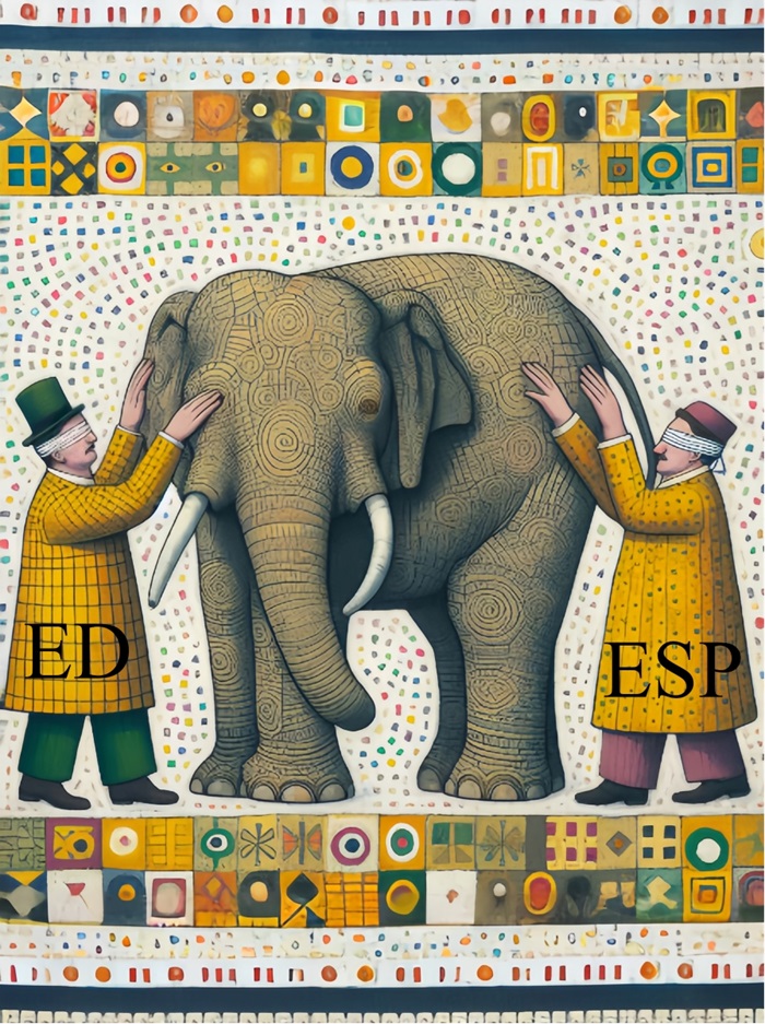
For example, Electron density (ED) is measured using X-rays, and electrons make it possible to measure electrostatic potential (ESP). Neutrons are sensitive to nuclear coherent scattering length (NCSL).
For electrons as probes, reaching atomic resolution is a fairly new development dubbed the 'resolution revolution'. Interestingly, it was not entirely clear what kind of image (map) exactly one 'should' see with electrons at high resolution, even for already 'known' structures.
Bochtler was inspired by the cryo-electron microscopy (cryo-EM) technique, which allowed the groups of Professor Holger Stark and Dr. Ashwin Chari to obtain the first atomic resolution maps. These maps showed the unexpected effects of using an electron beam for imaging. Dr. Bochtler re-analysed these maps. 'Electron density maps measured using X-rays as the probe, and electrostatic potential maps measured using electrons as the probe, are widely regarded as equivalent except for experimental errors. Our findings show that this is not entirely the case’, he says.
'The resolution revolution in cryo-EM offers exciting opportunities. To take maximal advantage of it, we should fully understand what the maps actually show', Bochtler adds.
Using advanced theoretical models like density functional theory (DFT) and the Bethe-Mott relation, Dr. Bochtler has demonstrated that ED and ESP maps differ significantly in their depiction of atoms, particularly in how they display the charges within atoms. He has also shown that the relative contributions from lighter and heavier atoms, the sensitivity to chemical bonding in outer atomic shells, and the sensitivity to charge, all depend on the type of probe.
Dr. Bochtler's research not only adds to the theoretical foundations of atomic imaging but also enhances our ability to use cryo-EM for practical applications, including drug design.
His analysis indicates that X-rays, electrons and neutrons are each tuned to different aspects of matter, and thus a source of complementary information. X-rays detect electron density, while electrons used in cryo-EM sense the electrostatic potential, and neutrons are sensitive to nuclear coherent scattering length (NCSL).
'The resolution revolution in cryo-EM offers exciting opportunities. To take maximal advantage of it, we should fully understand what the maps actually show', says Bochtler.
Colleagues have now come out in praise of his work, saying: ‘Dr. Bochtler’s innovative study published in Structure provides a perfect example of how biological sciences are an integral part of STEM (Science, Technology, Engineering, and Mathematics), intertwined in the common quest to understanding the foundations of the world we live in’.
PAP - Science in Poland
lt/ zan/ kap/
tr. RL