Photosensitive cells in the eye - cones and rods - change their sizes for a moment when light flashes on them. Polish researchers have just explained why. This tiny, fast pulsation of retinal cells can be seen live thanks to the Polish method of eye imaging.
Professor Maciej Wojtkowski from the International Centre for Translational Eye Research (ICTER) at the Institute of Physical Chemistry PAS talks about this breakthrough discovery for eye research in an interview with PAP on the occasion of Polish Science Day.
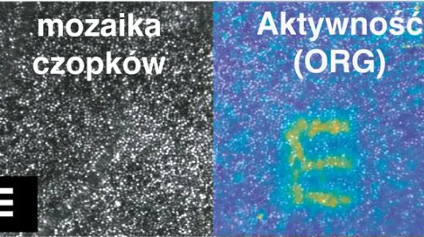
The retina contains photosensitive cells - cones and rods. When light flashes - for example in the shape of the letter E - photons from this character reach the photosensitive cells of the retina. These cells shrink for a moment, but only in those parts of the retina that light has reached. So when you look from the side at this part of the eye with great magnification, you can see the same shape there for a moment - in this example the letter E - emerging from the background of retinal cells. This means it is possible to see the image recorded by the eye - live. Now, a Polish team of scientists has explained what causes this effect at the protein level.

The research of the ICTER centre, founded and headed by Professor Maciej Wojtkowski, was published in the prestigious scientific journal PNAS. This is another step towards introducing the optoretinography (ORG) method developed by the Polish team to the market. It will be a non-invasive, non-contact, fast and objective method of eye diagnosis and detection of eye diseases at an early stage. It is based on recording changes in the length of retinal cells under the influence of fast flashes of light.
In the publication in PNAS , scientists show experimentally that the pulsation of retinal cells results from changes in the size of a certain protein - PDE6. It takes part in a cascade of changes that occur when the cell absorbs light.
As luck would have it, this protein reacts to the presence of sildenafil - known under the trade name Viagra - and therefore a well-studied active compound. This pharmaceutical, most often used in erectile dysfunction, ultimately acts on another protein from the same family (PDE5), but also blocks the PDE6 protein.
'A man once tried to commit suicide by overdosing on Viagra. He survived, but he developed permanent vision problems', Wojtkowski describes. It was already known that this potency drug in large doses affected vision, but it was not known how. New research sheds more light on this.
MICE ON VIAGRA LOSE AND REGAIN VISION
As part of Polish research, an overdose of sildenafil was induced in laboratory animals. As a result, for a certain period, PDE6 proteins in their retina stopped working, i.e. changing shape. It turned out that because of this, the animals went blind for a while. 'This was known, for example, from the analysis of the work of the visual cortex of the brain', Wojtkowski explains. When the effect of the pharmaceutical wore off, the visual cells regained their ability to lengthen and shrink, and the animals regained their vision. In this way, Polish researchers have shown that the changes in the size of cones and rods related to the work of the PDE6 protein under the influence of light are actually necessary for seeing.
PROTEIN LAYUP
It is impossible to observe changes in the shapes of individual proteins as part of their work live. However, nature used a certain trick in the eye. And Polish researchers also used it in their solution.
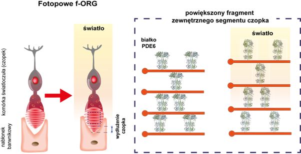
PDE6 proteins are arranged in levels in the outer layer of photosensitive cones. They fill subsequent levels of the elastic cell membrane folded thousands of times. Under the influence of light, a cascade of changes occurs in the cell. And then the proteins on each level of the folded membrane change their shape - they alternately slightly expand and contract their levels. Each level changes height by only a tiny bit - the size of an atom. However, the thousand-fold sum of such tiny bits has significance - it reaches tens or even hundreds of nanometers. And Polish researchers observe this thanks to their instruments in the living eye. The activity of PDE 6 proteins has a well-known key significance in the process of vision, so the observed change in cell length is not a coincidence.
A FLASH IN THE EYE
'In optoretinography, we shine short flashes on the eye. And we observe these very small - nanometer-sized - changes in the length of photoreceptors on the retina. At the time of the examination, we can see whether the photoreceptors in the eye respond to our signal correctly or not, and whether there are any irregularities in the work of the retina', the scientist explains.
In order to collect information about changes in the length of photosensitive cells, scientists had to develop a completely new imaging method called Spatial-Temporal Optical Coherence Tomography (STOC-T). This method is based on the specific properties of light reflected from the retina. In addition to the frequency and amplitude of changes in light reflected from the eye, it also analyses changes in the phase of this light. Thanks to this, it can record even nanometer changes on the surface of the eye between flashes. 'So we see what the eye sees', Professor Maciej Wojtkowski describes. The precision of these observations is well above that of previous methods.
Biochemical processes related to vision take place in a very small space - in the retina of the eye, and in a short time - several dozen times per second. What's more, they are very small, measured in nanometers. However, Professor Maciej Wojtkowski and his team came up with an idea for recording these small changes. Now, scientists from ICTER are taking further steps to better understand the mechanisms occurring in healthy and diseased eyes. All this knowledge is essential if we want to diagnose eye diseases better, detect changes in the early stages of the disease and stop them.
ICTER will implement one of three Polish research and innovation projects that received funding in the prestigious European Teaming for Excellence programme. Their inauguration took place in Warsaw.
PAP - Science in Poland, Ludwika Tomala
lt/ agt/