Professor Paweł Moskal from the Jagiellonian University has been awarded a prestigious European Research Council (ERC) Advanced Grant for a groundbreaking project that aims to transform cancer diagnostics. His team is exploring whether quantum measurements from a PET scan could non-invasively determine tissue oxygenation levels—a key indicator of tumour malignancy—offering a potential alternative to traditional biopsies.
“Our hypothesis assumes that tissue oxygenation can be assessed by measuring the degree of quantum entanglement of photons created during positronium annihilation. Theoretically, there are grounds to believe so,” said Moskal.
More importantly, it enables previously inaccessible quantum measurements, including the properties of positronium atoms—exotic matter formed when electrons and positrons briefly bind before annihilation.
More importantly, it enables previously inaccessible quantum measurements, including the properties of positronium atoms—exotic matter formed when electrons and positrons briefly bind before annihilation.
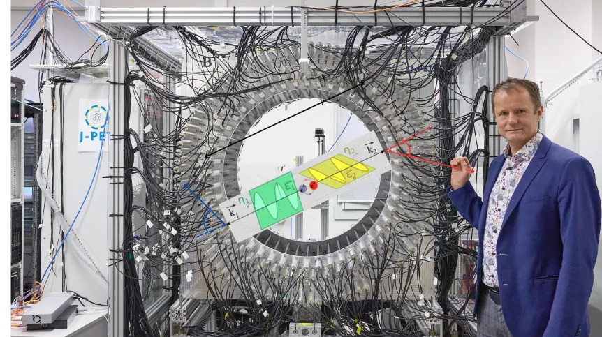
“We have the only tomograph in the world that allows to measure the degree of quantum entanglement of photons created from positronium,” Moskal said. “Thanks to this, we are the first in the world to discover and publish—in the journal Science Advances—that the degree of entanglement depends on the material in which positronium annihilates. Will such a measurement allow to assess tissue oxygenation? No one has tested this yet. We are taking this risk.”
Oxygen deficiency (hypoxia) is a known characteristic of malignant tumours. Rapidly dividing cancer cells consume oxygen intensively, while abnormal blood vessel growth in tumours exacerbates this by limiting oxygen delivery.
“If it were possible to determine which tissues are hypoxic using PET, it would be possible to non-invasively scan the entire body and identify areas where malignant tumours are developing—something like a biopsy covering the entire body,” Moskal said.
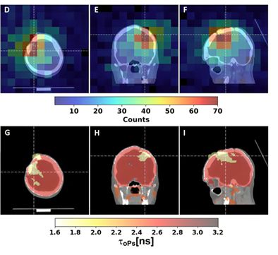
This concept, dubbed a “quantum biopsy,” could revolutionise oncology by enabling physicians to assess malignancy without physically removing tissue. It builds on earlier achievements: in 2024, Moskal’s team used J-PET to image a glioma in a human brain based on positronium lifetime measurements, a parameter not captured by traditional PET.
"Now, the team is pushing further by examining quantum entanglement properties of the annihilation photons. By analysing the polarization and coherence of photon pairs, the researchers believe they can extract information about the microenvironment of annihilation—including the presence or absence of oxygen.
“Even if the hypothesis is not confirmed, scientists will gain: they will expand their knowledge of the physics of annihilation and improve J-PET. However, if it does prove true, it may revolutionise cancer diagnostics, giving humanity a new tool—a non-invasive, full ‘quantum biopsy’,” Moskal said.
In the latest round of ERC Advanced Grants, announced in June, 281 top researchers from across Europe were selected to receive a total of €721 million in funding. Moskal is one of four Polish scientists to receive an ERC grant in this cycle. Each project will receive approximately €2.5 million over five years.
With over 40 patents already to his name, Moskal has a proven track record in developing diagnostic technologies. The ERC grant will allow his team to continue pushing the frontier of medical imaging, combining high-energy physics, quantum information science, and clinical diagnostics in an unprecedented way.
PAP - Science in Poland, Ludwika Tomala
lt/ zan/ jpn/