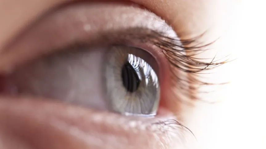
To be able to construct better eye implants, we need to know a lot more about the electrical signals that nerve cells send to each other. Polish researchers help make computer-to-brain communication even more precise.
Nerve cells use electrical signals to communicate with each other. Scientists have been trying to eavesdrop on this communication for a long time. Knowledge of brain waves - electrical activity of the brain - has been useful in medicine (EEG) and psychology (biofeedback). Understanding the brain waves is just the beginning - it is like understanding the pattern of electricity consumption by all residents of a borough. Scientists want to know how single "citizens" - individual nerve cells - use electricity. And they want to establish 2-way communication with them. This task is much more difficult.
EYESIGHT TO THE BLIND
The ability to stimulate nerve cells and to register their activity is crucial for the development of new generations of neuroprostheses. Over the past few years, implant surgery has been performed in the world, for example, to restore vision of people with retinal degeneration. Such a prosthesis is a device that converts camera signal into electrical pulses. These pulses are then sent to the retinal cells by means of electrodes. And then the nerve cells begin to send information about the image to the brain.
This could be hard to believe. But when we see, the information derived from the light is also converted into an electrical signal in the cells of the eye. If you know the characteristics of this electrical signal, you can reproduce this signal with devices and properly stimulate the specific nerve cells in the eye of the patient, thus giving him the ability to see.
For now, however, implants are not as good as our eyes - quality of the image they transfer is poor. Implants can help blind people find the door, but they will not see the door immediately, only after a moment. The brain needs time to interpret the imprecise signal from the prosthesis. More research is needed to make implants better. Such work is conducted, among others, by researchers at Stanford University, California. Researchers from the AGH University of Science and Technology in Kraków also participate in the project.
The aim of the project is to improve the efficiency and precision of retinal neuron stimulation. Researchers want to initiate activity similar to that of a healthy retina in these cells. To that end, scientists are testing different ways of stimulating neurons and record cell responses.
FAIRLY NERVOUS CELLS
Dr Paweł Hottowy from AGH described the device used in the research. It is composed of 512 tiny independent electrodes that are in contact with retinal nerve cells and transmit delicate pulses to the nerve cells (occupying a surface of 0.5 mm2). At the same time, the device collects information from the cells about their activity. This tells the scientists how the cells respond to the stimulation signal and whether the electrical impulse is sufficient to stimulate them. "This way we learn how to best stimulate the cells" - explained Dr Hottowy.
"Our role in the project is primarily to provide specialized electronic equipment for these studies" - said the physicist. He explained that this includes miniature integrated circuits that allow to supply precisely tuned electrical signals to the cell during the experiment, while simultaneously receiving, amplifying and recording weak signals produced in nerve cells. "Simultaneous stimulation and registration of neuronal activity is a particularly difficult task, and our measurement system is the only one in the world that allows to do that" - said the researcher.
The cooperation of Polish and American scientists is supported by the grant "Harmony" awarded to the team from AGH by the National Science Centre.
MAKE THE CELLS DO AS WE SAY
AGH graduate Beata Trzpil was also involved in the research on computer-nerve cells communication. As part of her MA thesis, the researcher developed software that allows the user to freely define the signals used to stimulate nerve cells.
"It is only through this software that it is possible to use the full functionality of the electronics used in the project" - said Trzpil. "Neurobiologists in California can now conduct research that was not possible before" - she commented. Her MA thesis was recently recognized as the best application paper at her university in 2016 and awarded the "AGH Diamond".
Beata Trzpil explained that in the experiments at Stanford University researchers use monkey eye retina - very similar to human retina. In vitro, such cells function only for a few hours after they are extracted. In addition, access to the retina of the monkey is difficult because the tissue is obtained from animals used in other experiments. Therefore, each experiment needs to be carefully planned to get as much information as possible about cell activity, and the reliability requirements of the measurement system are extremely high. The software developed by the AGH MA student was used in experiments while she was still writing her diploma thesis.
Dr. Hottowy says that he wants to use his knowledge acquired during the work with the American team in further research. Together with the Nencki Institute of Experimental Biology in Warsaw he prepares for research into the action of nerve cells in the brain. Since we now know how to provide information about the image to the brain, the logical next step is to consider how to supply other, equally complex information to neurons in the brain.
PAP - Science and Scholarship in Poland, Ludwika Tomala
lt/ agt/ kap/
tr. RL


