A 2,000-year-old Egyptian mummy that was thought to be pregnant, did not have a foetus in her pelvis but ‘a mummified organ’, say scientists.
Researchers at the National Museum in Warsaw said that despite X-ray scans and CT images last year revealing what appeared to be a foetus, this was the result of ‘a computer illusion and misinterpretation.’
Instead, the scientists say the images reveal four ‘bundles’.
In April 2021, part of the Warsaw Mummy Project (WMP) team published an article in the Journal of Archaeological Science, which shows that a woman's mummy belonging to the University of Warsaw and exhibited at the National Museum in Warsaw conceals a 7-month-old fetus inside. The authors of the publication came to this conclusion on the basis of the analysis of images obtained using X-ray and computed tomography. Not all researchers, however, agreed with this claim. It was questioned by i.a. radiology expert dealing with mummies - Prof. Sahar Saleem.
Writing in July’s Archeological and Anthropological Sciences (DOI: 10.1007/s12520-022-01598-z), bioarchaeologist and co-founder of the Warsaw Mummy Project Kamila Braulińska said: “The bundles were placed there by ancient embalmers.
“In the bundles there is probably at least one mummified organ of the deceased. It was a well-known practice in ancient Egypt.
“The remaining bundles may contain body parts or other products of the mummification process. There is also another possibility - embalmers placed bundles in the mummies in order to maintain the shape of the body after the mummification process.”
She added: “Our article contains a number of spectacular images and links to videos depicting the interior of the ancient mummy, including those made using holographic techniques, which are the latest trend in medicine.
“This is not the first mummy with bundles of this type. Objects of this type are sometimes found in other parts of the body, and similar bundles or substances are found in the pelvis.”
Because the body decomposes more easily when internal organs are not removed, ancient Egyptians instead removed them during the mummification process.
It was three of the four bundles in the pelvis that the previous team interpreted as a foetus.
That the woman was not pregnant is also evidenced by quantitative measurements of radiological densities, as well as the geometric arrangement of the bundles and the comparative characteristics of the materials inside the mummy.
According to the authors of the new article, their predecessors failed to consult a radiology expert prior to publication, although their conclusions were based on medical X-ray and CT images.
They add that the alleged discovery of the mummy's pregnancy resulted from an illusion caused by the phenomenon of pareidolia, a natural human desire to see familiar objects in random shapes.
Braulińska said: “This phenomenon, combined with the lack of consultation of theories with a radiology expert, unfortunately only brought the effect of a global sensation, and not a reliable scientific study.
“Our article proves how important the cooperation with specialists from various fields is in the study of ancient Egyptian mummies, and how rationally and critically one should approach the analysis of the results, putting illusions aside.
“The respect due to human remains is also important.”
The scientists say they used the same computed tomography data as the previous team and, among other things, the same software.
Radiologist Dr. Łukasz Kownacki, who performed a tomographic examination of the mummy and created its first 3D images, said: “We have shown how much the analysis of 3D effects and their interpretation depend on the skills of the software user, who can achieve excellent visualization effects also without being a radiologist.”
For the latest study, the researchers used the capabilities of radiological analyses available at the Department of Diagnostic Imaging of the European Health Centre Otwock, including unique medical holographic software for the Mixed Reality systems, as well as radiological server solutions.
In addition to Kamila Braulińska, the authors of the publication challenging the pregnancy of the mummy are Łukasz Kownacki, Dorota Ignatowicz-Woźniakowska (Chief Conservator of the National Museum in Warsaw and WMP coordinator for the National Museum in Warsaw) and Maria Kurpik (Senior Conservator of the National Museum in Warsaw, specialist in the field of conservation documentation, including imaging of works of art).
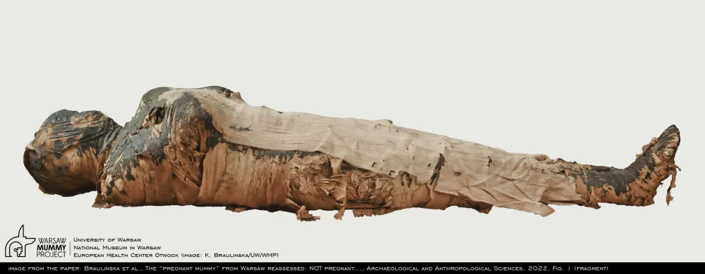
The mummy was brought to Poland in the 19th century. It belongs to the University of Warsaw, but is currently in a long-term deposit at the Ancient Art Gallery of the National Museum in Warsaw. (PAP)
author: Szymon Zdziebłowski
szz/ zan/ kap/
tr. RL
Gallery (8 images)
-
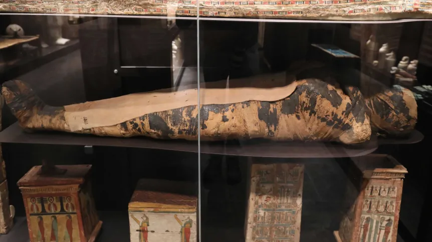
 1/8Egyptian mummy of a woman from the collection of the University of Warsaw, exhibited in the Gallery of Antiquities of the NMW. Originally, it was considered to be the mummy of a man, the priest Hor-Djehuti; this has been ruled out by research carried out by the Warsaw Mummy Project. K. Braulińska (UW/WMP)
 -
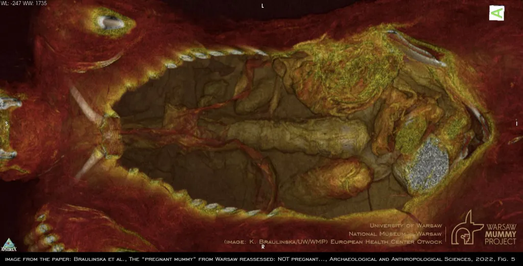
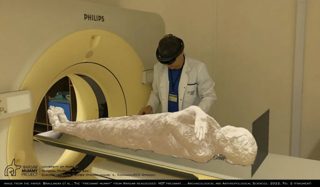 2/8A hologram of the mummy created using the mixed reality (XR) technique from computed tomography images. It was obtained using the Polish CarnaLife Holo software (MedApp) for HoloLens systems (Microsoft) and virtually placed on the tomograph table where the mummy was originally examined. Ł. Kownacki (ECZ Otwock)
 -
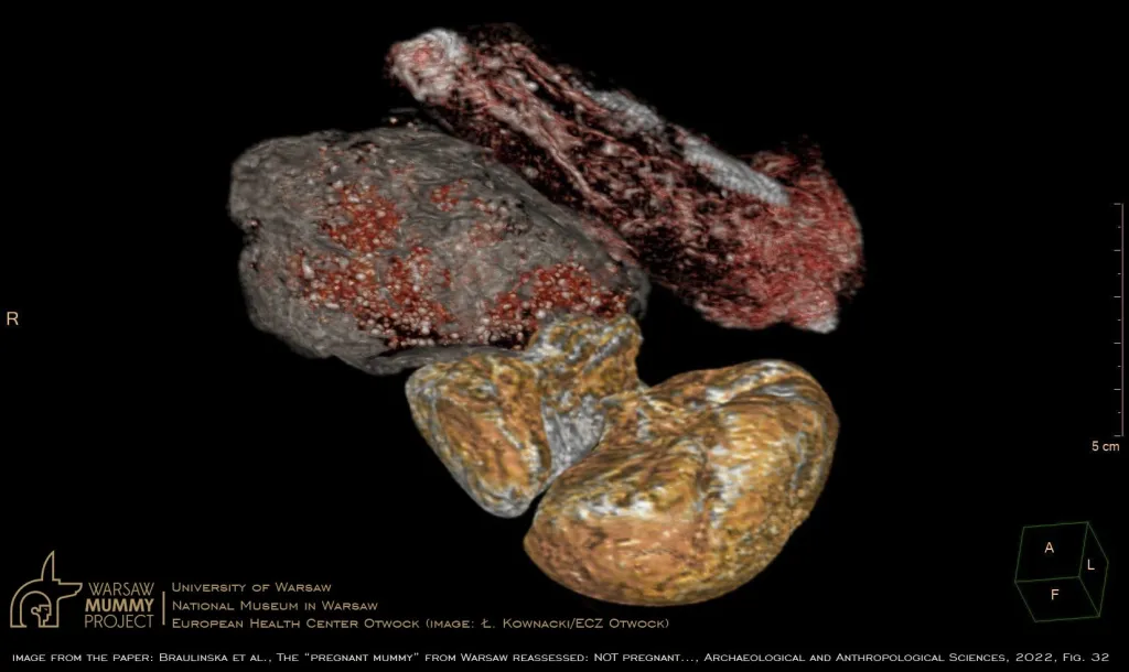
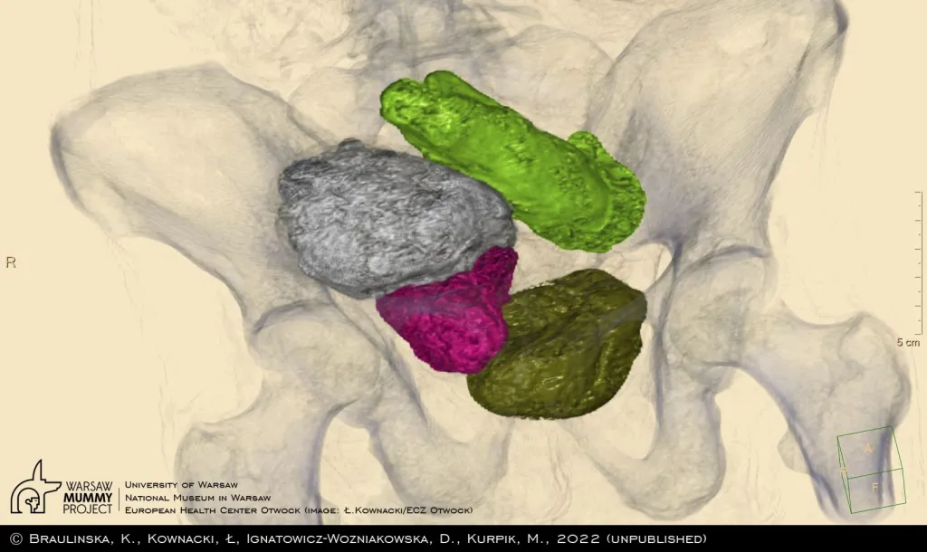 3/8The contents of the mummy's pelvis (front view), as a result of the segmentation of images, showing various types of packages placed by ancient embalmers, which (except for the upper one, which was omitted) were interpreted by the previous authors as the fetus. Image created with an IntelliSpace Portal (Philips) server. Ł. Kownacki (ECZ Otwock)
 -
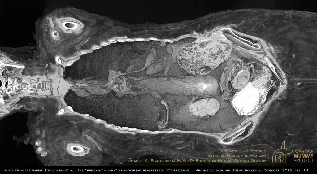 4/83D monochrome cross-sectional reconstruction of the mummy using OsiriX MD (Pixmeo) software. A view from the front to the insides of the mummy filled with packages of various nature, some containing internal organs removed earlier and mummified separately. K. Braulińska (UW/WMP)
 -
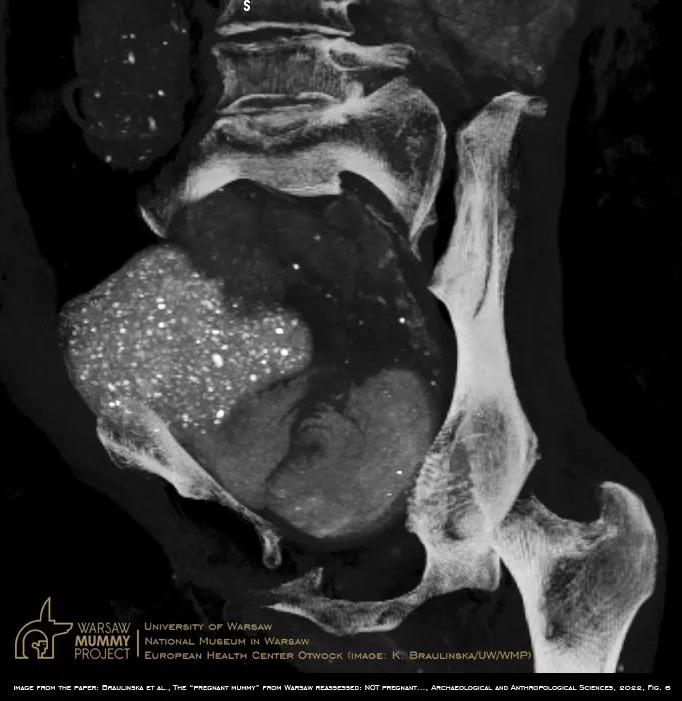
 5/83D reconstruction of separate packages present in the pelvic cavity of the mummy presenting diverse materials. The three lower packages considered as one were indicated by previous authors as a fetus. However, the largest of them, filled with a granular and dense heterogeneous substance, is certainly not the “head of a fetus”. Ł. Kownacki (ECZ Otwock)
 -
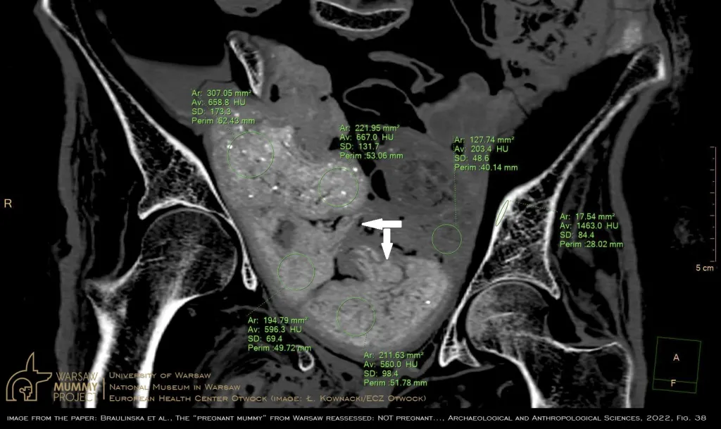 6/8Cross-section through the pelvis of the mummy (frontal view) shows different densities of substances filling it. Despite the lack of any skeletal and anatomical structures, the arrangement of packages and most likely the loose ends of the bundles of material appeared to the “discoverers of the pregnancy” (based on what they reported) as the “hands and legs of a fetus” (arrows). The cross-section through the largest package (“head”) reveals the granularity and high density of its material. Ł. Kownacki
 -
 7/83D color reconstruction of a cross-section through the mummy, using OsiriX MD (Pixmeo) software. Frontal view into the mummy filled with packages of different nature. The packages are partly immersed in resin, thus fixed in the different cavities of the mummy. K.Braulińska (UW/WMP)
 -
 8/8Reconstruction of the area of the alleged “fetal head” using OsiriX MD (Pixmeo) software. The visible effect of a “snowstorm” caused by numerous grains inside the largest package. These granularities have much higher radiological density than the bones of the mummified woman. Together with the absence of any anatomical structures typical of the skull, it completely excludes the possibility of a “fetal head” presence . K. Braulinska (UW/WMP)