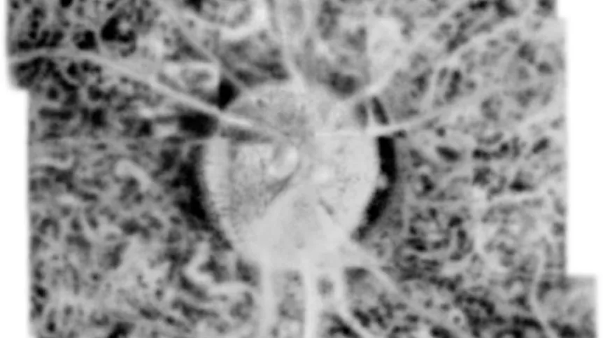
Polish scientists have developed a technique that enables visualization of the back layers of the eye - the retina and choroid - at discrete depths.
Called STOC-T, the technique has now been hailed by the Institute of Physical Chemistry of the Polish Academy of Sciences as “another milestone in eye imaging.”
Modern imaging of eye tissues would not be possible without Optical Coherence Tomography (OCT) scans. As one of the world's most popular and accurate diagnostic techniques, this method has enabled us to more fully understand the mechanisms of many diseases and select more effective therapies. However, OCT is not a perfect technique. Coherent noise and limited axial range have prevented high-resolution imaging, as well as full penetration of all layers of the retina and choroid.
Researchers at the International Centre for Translational Eye Research (ICTER) found a way around these limitations and developed Spatio-Temporal Optical Coherence Tomography (STOC-T). The latest research by the team led by Professor Maciej Wojtkowski confirms that this method makes it possible to view the retina and choroid with high resolution at distinct depths in the frontal section.
According to the Institute of Physical chemistry, no one in the world has previously succeeded in doing this.
In a previous paper published in Biomedical Optics Express, ICTER researchers described their STOC-T time-frequency OCT system for capturing retinal optoretinograms.
Now, Professor Maciej Wojtkowski's team at ICTER, in a paper published in iScience (https://doi.org/10.1016/j.isci.2022.105513), has shown that retinal images obtained with STOC-T maintain a uniform resolution of ca. 5 μm in all three dimensions, across a thickness of about 800 μm. This, in turn, makes it possible to obtain high-contrast, volumetric images of the choriocapillaris with reduced scattering effects.
Professor Maciej Wojtkowski from ICTER said: “We applied known data processing algorithms and developed new ones to handle and process the acquired data sets to obtain high-contrast 3D data (volumes) for the retina in large fields of view. The technology and algorithms made it possible to image the retina and choroid at high transverse resolution at different depths, making the differentiation of morphology visible for the first time within the Sattler, Haller, and choriocapillaris layers.”
The main limitation of this breakthrough method is the current price of the camera that costs around 100,000 euros. The scientists expect that as the volume of camera production increases, the cost would gradually drop.
STOC tomography enables distinct imaging of all primary layers of the choroid while showing difficult-to-image layers over a large transverse and axial range. The data can only be analysed offline due to the low transmission speed between the camera and the computer. Considerable computing power is required to process all the vast amounts of data, but this can be reduced by using machine learning algorithms such as deep learning.
Using STOC-T for retinal imaging allows to reconstruct the morphology of the cones of the human eye. With the aforementioned camera, the STOC-T method makes it possible to capture the retina in a fraction of a second and record its entire depth in extremely high, unprecedented resolution, assure representatives of the Institute of Physical Chemistry PAS.
In clinical practice this means that the eye will be fully imaged with an accuracy that allows single cells to be viewed before the patient has time to blink. 'STOC tomography has the potential to usher in a new era in the diagnosis of eye diseases, although much more practical refinement needs to be done before it can be routinely disseminated', conclude the representatives of the institute.
Find out more about the solution on the Institute of Physical Chemistry PAS website.
PAP - Science in Poland
lt/ zan/ kap/
tr. RL


