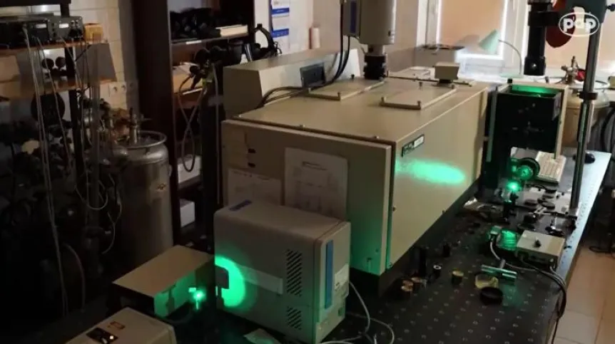
Is it possible to detect cancer and determine its malignancy degree in a few minutes or even seconds? It turns out that it is. This is possible with innovative diagnostic tools: Raman optical biopsy and virtual histopathology, developed in the Laboratory of Laser Molecular Spectroscopy (LLSM) of the Lodz University of Technology.
According to the researchers, this is a qualitative breakthrough for patients, as it enables oncologists to receive a precise, objective test result in real time.
Head of the Laboratory of Laser Molecular Spectroscopy Prof. Halina Abramczyk emphasizes that the laboratory has developed a commercialisation-ready, innovative optical biopsy method for the identification of tumours and virtual histopathological analysis, based on measurements of Raman scattered light. Researchers have also developed a Raman surgical navigation method that facilitates tumour removal during surgery.
"We also have tests that will allow to detect Raman tumour markers based on a blood test in seconds" - adds Prof. Abramczyk.
Researchers say that innovative technology based on Raman spectroscopy used to identify cancer and create images of tumour tissues is completely safe for the patient.
The test does not require extracting tissues from the body. It involves illumination of the examined tissue suspected of the presence of neoplastic lesions with laser light using a fibre optic probe, and the analysis of Raman spectrum generated in seconds as a tissue response.
Researchers from the Laboratory of Laser Molecular Spectroscopy focused on four types of cancer: breast, head and neck, gastrointestinal tract and brain cancer.
"Our innovative method is not only a modern technique based on the phenomenon of Raman light scattering, but also a method of finding biomarkers. It took us about 10 years. We have a database for tissues of about 300 patients that contains hundreds of thousands of spectra. We are able to determine the degree of malignancy of the tumour in seconds, and in some cases with imaging - in minutes" - adds Prof. Abramczyk.
Dr. Jakub Surmacki from the Laboratory of Laser Molecular Spectroscopy emphasises that the Raman method of surgical navigation developed by the researchers from Łódź enables the unambiguous determination of the margin of error during surgery. "This is extremely important during the procedure, so that the surgeon knows whether he has completely removed the tumour" - he says. Raman optical biopsy allows for quick and unequivocal identification of the cancer and its advancement degree.
"We introduce a biopsy needle into the breast affected by the tumour. We attach a fibre optic probe with laser light to the needle and in a few seconds we are able to register the Raman spectrum on the computer screen. We get a clear information, in real time, about the degree of malignancy of breast cancer" - explains the scientist.
Another invention developed in the laboratory is virtual Raman histopathology, which uses the same phenomenon of light scattering. Researchers` results have proven that it allows to obtain identical results as standard histopathology based on tissue morphology analysis that uses hematocyllin and ozone staining.
However, according to the invention co-author Prof. Beata Brożek-Płusk, a huge advantage of the Raman virtual histopathology is the fact that the results are obtained in minutes. "We radically shorten the doctor`s, and consequently also the patient`s waiting time for the diagnosis" - says Prof. Brozek-Płuska.
The researcher also emphasises that Raman imaging, which is used in this method, can also be used to look inside the human body, which was unavailable in other methods. It allows, for example, to image human milk ducts.
The researcher presents a comparison of the results for a milk duct with a normal structure and for a duct affected by cancer.
"In the case of a healthy duct, its interior is empty, but thanks to Raman imaging we are able to visualize subsequent structures that occur around the light of this duct. In the case of a duct, in which tumour develops, we are able to visualize cancer cells present inside, as well as examine the structures that are around the duct and thoroughly determine the biochemical composition of the examined structures" - emphasises the invention co-author.
According to the researchers, laboratory tests carried out at the Laboratory of Laser Molecular Spectroscopy on tissue preparations taken from several hundred cancer patients clearly demonstrate that the developed innovative procedure of optical biopsy and virtual histopathology is fast and objective, because the test result is based on the bands recorded in the Raman spectrum; it is independent of interpretation and experience of medical personnel. The method is also extremely sensitive - at the level of 90 percent.
In addition, tissue examination is possible without the use of contrast. This technique also allows to estimate the degree of histological malignancy of the tumour, and the identification of neoplastic lesions occurs with the precision of a fraction of a micrometer.
The researchers from Łódź emphasise that currently they are primarily interested in introducing this method in the Polish economy and medicine as soon as possible. "The more so because the first paper describing surgery using Raman neuronavigation carried out in the United States, appeared in January. Unfortunately, in this situation, we will not be the first in the world, but we can be the first in the country and in Europe" - concludes Prof. Halina Abramczyk.
PAP - Science in Poland, Kamil Szubański
szu/ agt/ kap/
tr. RL


