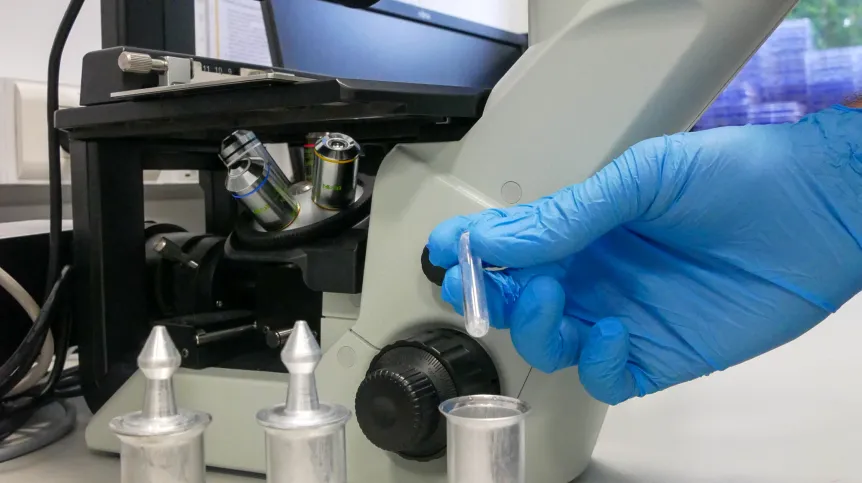
In a few years, the solutions to fight liver cancer may include yttrium oxide gel microspheres produced at the National Centre for Nuclear Research in Świerk.
In a press release the institute said that the results of the latest research on the impact of new microspheres on biological material are promising.
“If further tests confirm them, in the future microspheres can be produced quickly and cheaply, precisely matching their characteristics to the needs of a particular patient,” the press release said.
Malignant liver tumours are dangerous diseases with a high mortality rate. One of the promising methods of their treatment is radioembolisation. During the treatment, millions of radioactive microspheres with a carefully selected diameter and activity are introduced into the tumour tissue. When they get stuck in narrowing arteries, they cut off the tumour's supply of nutrients. At the same time, for a strictly defined period of time, their radiation kills sensitive cancer cells in the immediate vicinity.
There is increasing evidence that in the future these microspheres can be manufactured quickly and cheaply to meet the therapeutic needs and anatomy of specific patients. These conclusions are drawn from experiments on new microspheres with the yttrium 90Y isotope, produced and tested by a team of employees of Warsaw scientific and medical institutes with a significant contribution of the National Centre for Nuclear Research (NCBJ) in Świerk.
Research project leader, Dr. Edward Iller at NCBJ's Radioisotope Centre POLATOM said: “The microspheres we have been dealing with for several years contain yttrium oxide. Yttrium, after irradiation with neutrons in the reactor, is converted into the radioactive isotope 90Y with a half-life of 64 hours. In practice, this means that each microsphere, after being introduced into the body for ten days, would interact with the electrons it emits on cancer cells located at an average distance of about three millimetres.”
The new micrograins with spherical shapes are produced by the sol-gel method. It is a complex, multi-stage process, improved several years ago to match the requirements of radioembolisation of the liver by scientists from POLATOM and the Institute of Nuclear Chemistry and Technology in Warsaw. The first step is to prepare a colloidal solution - a sol - containing compounds-precursors of the target particles, including yttrium oxide. The sol is then properly coagulated, after which the solvent is removed and the sol is converted to a gel. After drying, the gel turns into a powder that stabilizes at high temperature. The process is fully controlled and enables the production of microspheres with the desired diameters ranging from 20 to 100 micrometers.
“In the case of our microspheres, we could individualize the therapy by selecting the activity of the microspheres in terms of the anatomical structure of the liver and tumour in a particular patient. However, before the first clinical trials take place, we need to conduct detailed studies on the effects of our microspheres on cells and tissues in the body. We have just finished the first series,” said Maciej Maciak from the Department of Radiological Metrology and Biomedical Physics of the National Centre for Nuclear Research, the first author of the paper published in the journal Radiation Physics and Chemistry (https://doi.org/10.1016/j.radphyschem.2022.110506).
In the latest tests financed by the National Science Centre, the researchers examined the physical, radiometric, dosimetric and biological properties of the microspheres. They established that after irradiation with neutrons, high concentrations of the radioactive isotope 90Y were present in the samples.
In the series of studies, experiments with biological material were of particular importance. Research topics related to the use of microspheres with yttrium oxide for radioembolisation of the liver have been developed from the beginning in close cooperation with radiologists from the Military Institute of Medicine in Warsaw. It was there that in vitro experiments verified the effects of microsphere radiation on the number of colorectal cancer cells.
In accordance with the predictions and simulations, it was observed that the number of cancer cells decreased with the increase in the number of microspheres interacting with them. In addition, researchers at the Warsaw University of Life Sciences performed histopathological examinations on an animal model. They established that the microspheres were located in blood vessels as expected.
The scientists said: “The results of all experiments confirm that the new yttrium oxide microspheres are a promising alternative to currently available commercial agents and qualify for further biological and preclinical studies.” Before that happens, though, the team involved in the project is planning to refine the microsphere manufacturing technology so that it is ready for potential applications beyond the laboratory scale.
PAP - Science in Poland
lt/ agt/ kap/
tr. RL


