Scientists from the Warsaw University of Technology have fed artificial intelligence with ECG data to enable doctors to diagnose heart attacks quicker.
Physicist Dr. Teodor Buchner from the university told PAP: 'Our method of imaging the ECG result shows the actual condition of the muscle, which will allow the doctor to diagnose heart attacks and arrhythmias faster and more effectively.’
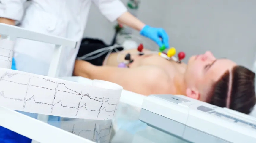
Electrocardiography (ECG) is a diagnostic test used in medicine to diagnose heart disease. It records the electrical activity of the heart muscle from the surface of the chest. The measurement result is a potential (voltage) difference between two electrodes, presented graphically on special graph paper or on a screen.
The ECG is one of the most difficult tests to analyse in medical diagnostics, and cardiologists have been struggling for years with problems in assessing the condition of patients based on the information they obtain from ECG. A large part of heart attacks are not even registered by the ECG test, while being able to react fast is critical for saving a person's life. An infarction that is 'invisible' for the ECG can be detected only after the patient reaches the hospital, where additional tests are be performed. Reducing the time needed for diagnosis and treatment would help to reduce mortality.
Another problem in cardiology is the localization of the disorder. It is not always clear whether the event - such as the source of an arrhythmia or an infarction - is located in the left ventricle, in the right ventricle, or perhaps in the interventricular septum. Based on the surface ECG, it is still very difficult to tell where exactly in the muscle something has happened, says Professor Rafał Baranowski from the Institute of Cardiology in Anin, one of the clinicians participating in the study.
'Previously, our team dealt with the heart from the +control system+ side: heart rhythm and its changes, so we already knew a bit about it. As to what happens to the muscle itself, we investigated it with electrophysiologists such as the unforgettable professor Franciszek Walczak, thanks to which we had an idea of the electrical activity of the heart. We have recently become interested how to interpret the ECG, and why it is described the way it is,’ says Dr. Buchner.
Buchner's team has applied for a patent for a solution that consists in improving the ECG with the help of artificial intelligence. 'We have formulated a theory that simply links the information from the ECG test result with information about the condition of the muscle. We believe that our way of imaging the ECG result shows the actual condition of the muscle. We show where the signal visible in the ECG physically came from and how it can be interpreted. A neural network is used for this analysis, so we are part of the search for new tools using the possibilities offered by AI,' he says.
He also explains what information artificial intelligence processes: 'It processes the ECG signal and outputs information that directly refers to the condition of the muscle. Currently, you have to guess based on the ECG graph that some wave on the graph is 2 mm and another is 3 mm - and what that actually means. There are diseases so difficult to diagnose that more than 70 parameters have already been defined to describe the ECG and there is still no consensus on which ones are the best. Thanks to our solution, artificial intelligence translates this abstract picture - into an image that is easily understood by the doctor.’
When asked about the possibility of a mistake made by artificial intelligence and the risk of misleading the doctor, he says: 'We, as engineers, are very critical of artificial intelligence, because we know that no algorithm is more intelligent than the human who wrote it. And that AI inherits all our limitations. We can order it to see something we haven't seen, but it still takes place in an +intellectual sandbox+ defined by a human, and the algorithm only executes human commands.'
Is artificial intelligence capable of finding connections between what is shown on the graph - and the condition of the heart muscle, connections the doctor does not see? 'We say: dig in this sandbox for me and take out all the pebbles. But we set up this sandbox, that is, we provide the data. And we know each of these pebbles, because we define the language in which the solutions are described. Artificial intelligence scales our efforts better. It will do it very quickly,’ Buchner says.
Now that the algorithm will combine information about the muscle with what the ECG test result shows, it can be assumed that this will weaken the vigilance of the doctor when interpreting the results.
To avoid the risk of weakening the cardiologist's vigilance, Buchner's team developed an educational tool. 'We have created an educational tool that shows what physically - in connection with the work of the heart - is the source of what the doctor sees on the ECG chart. Thanks to this tool, doctors can develop a better intuition, because so far they have been analysing the appearance of the ECG curve. It is like trying to read the pattern on a stocking when it's curled up into a ball,’ Buchner says. He emphasizes that doctors have great intuitions, so it is worth providing them with tools that develop their imagination to analyse difficult cases.
'This will result in a faster and more precise diagnosis, so the clinical path may change. It may happen that the patient will receive treatment already in the ambulance, because the doctor will have a greater degree of certainty about what is happening to the patient,’ says Buchner.
When asked about the databases fed to the artificial intelligence, the scientist says he hopes that the existing databases of patients' ECG results will be made widely available. 'The databases are now dispersed, and we believe that they will be integrated for the benefit of humanity. India already has a huge database of a million ECG test results,’ he says.
PAP - Science in Poland, Urszula Kaczorowska
uka/ zan/ kap/
tr. RL

