A laser that emits short pulses, developed at the Wrocław University of Science and Technology, now helps in examinations of the retina and will be useful for analysing nanomaterials and activating chemical reactions. Scientists plan to commercialise the device that can potentially replace older tools of this type in science and research.
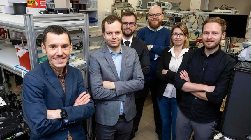
Dr. Grzegorz Soboń's team from the university’s Faculty of Electronics, Photonics and Microsystems, who developed the solution, recently received the Nikola Tesla Award presented by the Wrocław University of Science and Technology for outstanding scientific or engineering achievement.
'Our team has been dealing with lasers for many years. As part of one of the projects, we developed a pulsed fibre laser, which emits short +flashes+ of light. They appear at regular intervals and are extremely short. In this case, it means several dozen femtoseconds, where a femtosecond is one quadrillionth of a second,’ Dr. Soboń says.
IMPULSE SHORTER THAN THE BLINK OF AN EYE
The fibre laser is a device of a much simpler design than the femtosecond titanium-sapphire laser currently used to generate short pulses. The laser version designed by Wrocław University of Science and Technology experts is also a much easier to use, it does not require laser technology skills and can be operated by a doctor, biologist or chemist.
'We have prepared it so that you just press one button and the laser works. This is also very important in the case of implementing this solution,’ Dr. Soboń says.
Research on the interactions of such short pulses with matter opens the way to observing a variety of phenomena. Therefore, the possible applications of this laser include multiphoton microscopy for examining the structure of tissues, the production of nanostructures on surfaces, the study of modern nanomaterials or fluorescent proteins that are important for neurodegenerative diseases.
'It can be used to analyse various biological structures and look deep into tissues such as the brain. You can activate certain processes and chemical reactions, which may be interesting for chemists,’ the scientist explains.
SAFE LASER INTO THE RETINA
Currently, the laser developed at Wrocław University of Science and Technology is used for human retina examination and imaging at the International Center for Translational Eye Research (ICTER).
'The eye is a priceless and sensitive organ for humans, and any laser equipment that would be used to shine light into the eye must meet very stringent safety requirements. There were no lasers on the market with sufficient parameters to observe the effect we wanted when imaging the eye, that at the same time met safety standards,’ Soboń says.
This imaging technique was previously used on mice using titanium-sapphire lasers. According to Dr. Soboń, those structures are quite old, large, complicated and expensive, and their parameters were not suitable for safe examination of human eyesight.
The Wrocław scientists conducted the first tests of their laser on extracted biological samples. Later, when it turned out that the technique worked, they built a prototype laser, which was installed at the ICTER Institute. There, it was integrated with an ophthalmoscope, a device for examining the fundus of the eye. In 2022, the first tests on the human eye were carried out, and then a dozen more. The laser is still operated there, enabling research on mice and human vision.
'In the eye, in the process of vision, a cycle of chemical reactions takes place, triggered by light as a result of the impact of a photon on photoreceptors. At certain stages of the vision process, molecules are created that emit fluorescence. If we illuminate a given molecule with light of a certain colour, it will respond with a characteristic pattern of light of a different colour,’ says Dr. Soboń.
In the ophthalmoscope system, the patient's retina is excited by laser light and substances such as vitamin A metabolites emit fluorescence with a characteristic colour. Researchers record it and on this basis they receive information about the properties of the retina, e.g. whether any unfavourable changes are visible.
Experts at ICTER came up with this idea for retina examination. The problem with this type of examination was that the fluorophores would have to be illuminated with visually harmful ultraviolet light to fluoresce. Therefore, ICTER uses two-photon absorption, which circumvents the phototoxicity of UV light by using a fibre laser.
TRICK THE MOLECULES
'In this process, we trick the retina and illuminate it with light that has a wavelength twice as long, which is not phototoxic. Molecules that absorb two photons from this radiation +think+ they are excited by ultraviolet light, but they are not. In this way, we gain access to molecules that were not available before, because it was not possible to illuminate them in this way,’ Dr. Soboń adds.
The technology used in the described research is two-photon excited fluorescence microscopy.
The laser developed at Wrocław University of Science and Technology is quite universal and can be used for various applications. This is because the two-photon absorption effect is well known and used in many other branches of science - not necessarily related to the eye.
The researchers are currently concluding a project financed by the National Centre for Research and Development, in which they are developing a pre-commercial product - a solution that can be implemented at the highest level of technological readiness.
'We want to commercialise it. The assumption is that every laboratory that carries out retina or other research would be able to use our laser,’ Soboń says.
PAP - Science in Poland, Ewelina Krajczyńska-Wujec
ekr/ zan/ kap/
tr. RL


