
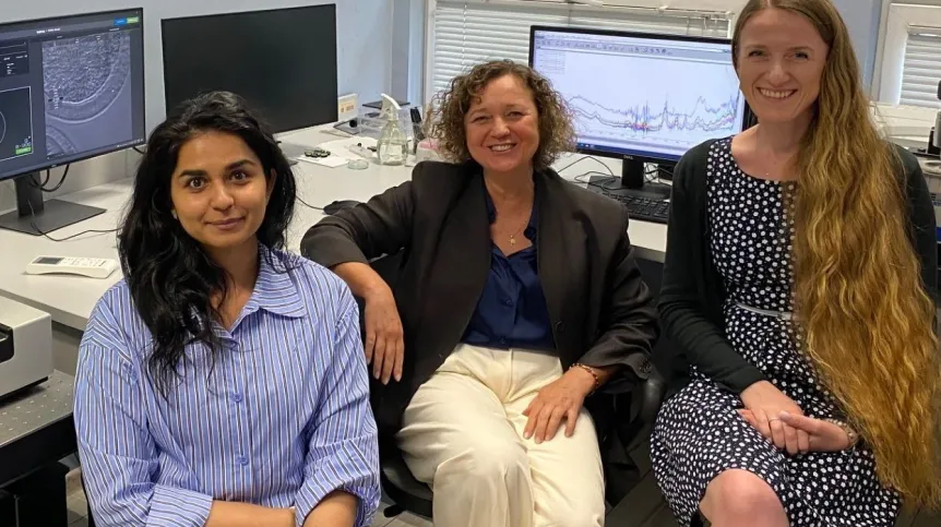
The distribution of lipid droplets inside an egg cell could reveal its chances of developing successfully, according to researchers from Kraków who have developed a non-invasive way to examine living human egg cells in three dimensions.
The researchers are the first in the world to use holotomography to examine living human egg cells in this way. Their method is designed to improve the selection of egg cells used in in vitro fertilisation (IVF) and increase the chances of successful implantation and the birth of healthy children.
The success rate of IVF averages around 35 percent. Because the rate is relatively low, extra embryos are created and stored so they can be implanted sequentially if previous attempts fail.
The concept developed by researchers from the Małopolska Centre of Biotechnology at Jagiellonian University and the Institute of Nuclear Physics of the Polish Academy of Sciences aims to reduce the number of embryos created while increasing the rate of successful implantations.
During IVF, a woman receives hormones that stimulate the simultaneous maturation of multiple egg cells. This can result in anything from a few to dozens of mature oocytes being retrieved in a single procedure. Only some are selected for fertilisation. The challenge is determining which egg has the greatest potential.
‘In every cycle, the body selects the single best oocyte, which is ready for fertilization immediately following natural ovulation. When a woman undergoes hormonal stimulation, we do not know which of the eggs would have been the one naturally selected. Under a light microscope, all the egg cells retrieved by the doctor look more or less the same’, Professor Grażyna Ptak of Jagiellonian University says in an interview with PAP. This raises the question of whether we can take a cue from nature and perform a precise selection.
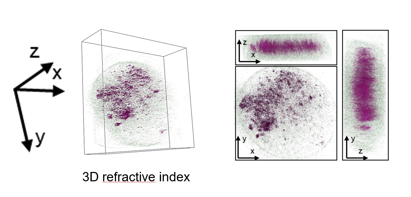
Researchers from Jagiellonian University and the Polish Academy of Sciences (PAN) see holotomography, a type of optical microscopy, as a promising solution.
‘Holotomography allows us to view a living cell in three dimensions without using any markers’, says Joanna Depciuch-Czarny, PhD, from the Institute of Nuclear Physics PAN, a co-developer of the new technology.
Holotomography relies on the fact that light refracts differently depending on the structure it passes through. For example, it behaves differently when passing through fat than through water. This allows structures and molecules inside individual living cells to be observed non-invasively during a procedure lasting about 10 minutes.
‘In our case, we focus on lipids; specifically, we observe lipid structures within the oocyte’, Ptak says.
For years, her team has investigated the role of lipid droplets in egg cells and embryos. The droplets store energy for the cell, act as a reservoir for signalling molecules and help protect the cell.
‘When something goes wrong within the cell, toxic oxidation products begin to accumulate in the lipid droplet. So, when we see large lipid droplets, it is a sign of reduced cell quality’, Ptak says.
In animal studies, her team observed a strong correlation between an altered lipid profile in oocytes and abnormalities in the resulting embryos, foetuses and adult offspring, particularly in lipid-rich tissues.
‘The egg cell gives rise to the organism. Abnormalities in the oocyte's lipid profile remain visible in the tissues of the resulting organism. Moreover, similar changes persist in subsequent generations of mice’, she says.
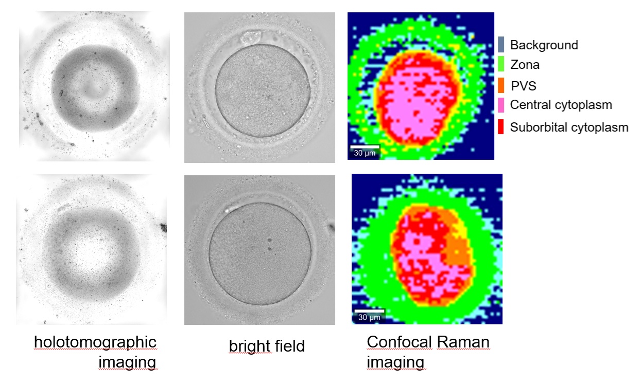
Until now, holotomography has primarily been used to assess small, adherent somatic cells. In their patent application, the researchers demonstrated how the technique could be used to assess the quality of a living, non-adherent oocyte.
This is a significant technical challenge because the egg cell is the largest cell in the human body and, being spherical, moves within the microscope's field of view.
‘Developing our method required solving numerous technical problems. One was finding an effective way to image a single cell suspended in culture medium using a holotomographic microscope. Another challenge was immobilizing that single cell, which does not adhere to a substrate. We are pioneers in this area’, says Hafsa Gulzar, a co-developer of the technology, quoted by the Jagiellonian University press release.
The researchers are now developing AI-based software that will automatically detect cellular pathologies from holotomographic images, allowing embryologists to identify abnormal oocytes more easily.
‘We expect the software component to be ready within a few months. Subsequently, collaboration with holotomographic microscope manufacturers will be necessary to create the software overlays required to perform these types of analyses’, Ptak says. She hopes IVF clinics will take an interest in her team's solution.
‘Our method allows to select cells with a normal lipid profile, which translates to greater procedural safety and a higher rate of healthy pregnancies. It offers a chance to reduce the creation of surplus embryos that are not transferred during a given cycle. If we can precisely identify the best cells, clinics and assisted reproduction centres could, for instance, fertilize only two selected oocytes instead of ten’, she says.
PAP - Science in Poland, Ludwika Tomala (PAP)
lt/ agt/
tr. RL