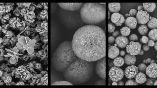
Polish scientists from three research centres are breeding genetically modified animals. Experiments on pigs and sheep gave excellent results. Researchers remove pigs’ antigens responsible for the proteins, against which the human body particularly strongly defends. Extracted tissues are used to make bioprosthetic heart valves.
The project "Transgenic Animals" was headed by Dr. Piotr Wilczek of the Foundation of Cardiac Surgery Development in Zabrze. National Research and Development Centre allocated 3.5 million zlotys to the project.
REDUCE THE INFLAMMATORY RESPONSE AFTER TRANSPLANTATION
Researchers from Zabrze, with colleagues from the Institute of Human Genetics PAS in Poznań and the Institute of Animal Production - National Research Institute in Balice, proved that genetic modifications allows to obtain very good biological material for valvular bioprostheses. Scientists are trying to prepare tissue of animal origin so that it assumes the appropriate functions in the human body and is not rejected.
This is yet another project led by Dr. Wilczek. It goes a step further than previous research into valves ready for transplant. More on previous work in PAP - Science and Scholarship in Poland here: http://naukawpolsce.pap.pl/aktualnosci/news,397263,wszczepic-trwalsze-zastawki-serca.html
The genetically modified animal tissue forms a matrix that is safe for the patient, a kind of scaffolding on which human cells then are then cultured. The pigs have the most critical antigens removed, to which the human body can react.
The team of Prof. Zdzisław Smorąg from Kraków bred dozens of animals with one of the critical antigens remove. The group of Prof. Ryszard Słomski from Poznań verified whether the modifications had taken place as planned. Laboratory tests in Zabrze were carried out to determine whether tissue engineering techniques work well in genetically modified animals. Previously developed methods concerned only non-modified animal tissues.
"There is always a risk that changing one gene can indirectly affect the other genes. We had to make sure that this modification did not affect any other feature that would determine the structure of the extracellular matrix, and that, after removal of cells, the tissue would not be weaker than the one in unmodified animals" - explained the project leader.
SWINE VALVE WORKS IN SHEEP
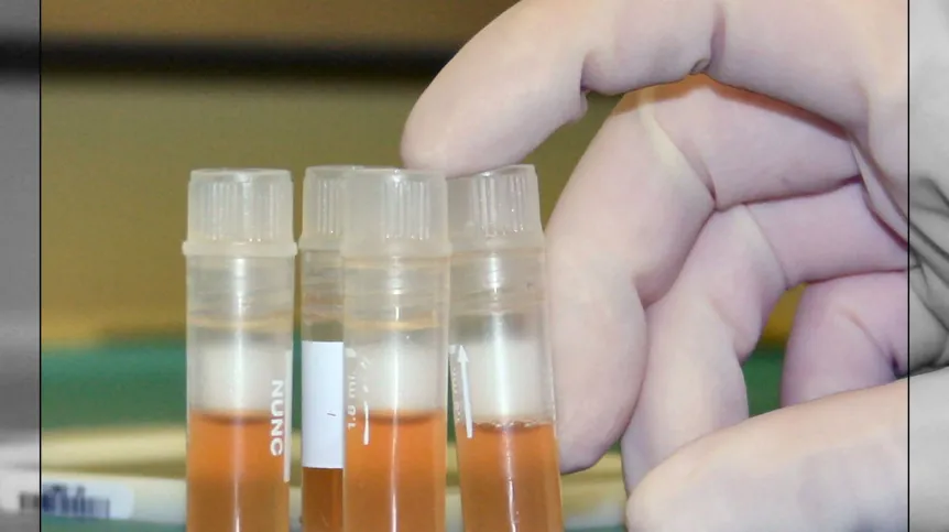
The main part of the work was the experiment on animals. The researchers wondered how the immunologically safer matrix would work in an animal model. The valves were built by covering the "scaffolding" of the porcine tissue with sheep cells. Then they implanted them in sheep, to see how the immune response associated with the presence of biological material derived from pigs can be suppressed.
"We used pulmonary valves. In patients, we usually replace their damaged valves with bioprosthetic valves. This was a bit different. Animals’ valves were left in place, and pulmonary valves implanted in the descending aorta. This was easier than pulmonary implantation, which would be surgically more complicated" - said Dr. Wilczek .
He explained that this complicated procedure requires entering extracorporeal circulation and contains many other threads that might distort the result of the experiment. It would not be known whether any failure would result from the bad valve, or the surgical technique.
"Load and pressure are higher in the descending aorta, so we knew that if our valve would work well in this environment, it would also properly function mechanically in the pulmonary position, and the risk of calcification would be limited" - explained the project leader.
The study was conducted on a group of 30 animals. In some sheep the researchers used commercial valves, in the other groups they implanted valves obtained in result of various modifications.
All animals had to carry the implanted tissue for a minimum of six months. The sheep survived and were healthy. After 8 months, scientists removed the valves and evaluated them in laboratory. They checked for any inflammatory infiltrates and calcification . According to Dr. Wilczek, calcification and calcium deposits make the valve unsuitable for further research.
"In the target group, where the valves were covered with sheep own cells, there was no calcification, and tissue looked good. I would venture to say that the image of the tissue was better than of the commercially available ones, which were used in the experiment" - summed up Dr. Wilczek.
PAP - Science and Scholarship in Poland, Karolina Olszewska
kol/ mrt/
tr. RL