Professor Krzysztof Palczewski and team are working on therapies for people with diseases that lead to loss of eyesight. At this point, gene editing techniques allow to repair genetic mutations in living cells, including the eye. 'We need 5-7 years before human trials could begin', he says.
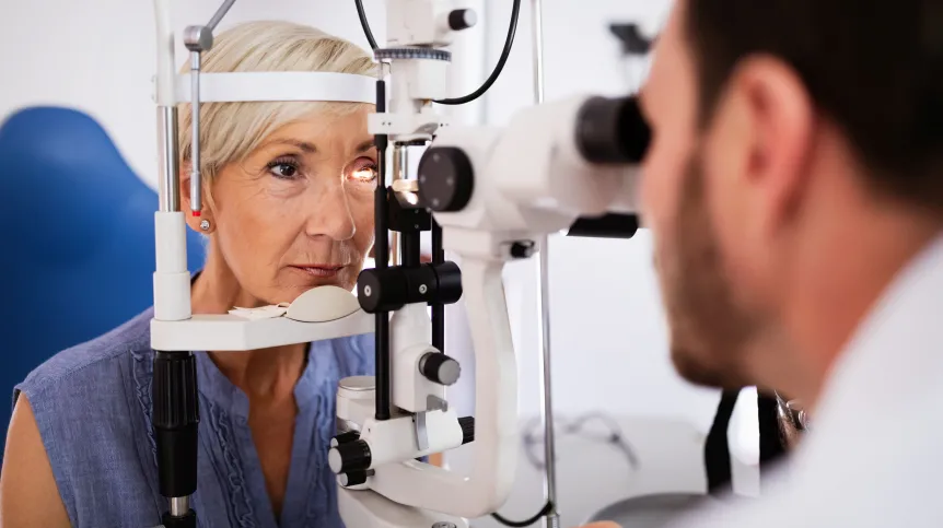
The procedure in question involves editing genes in the eyes of patients who suffer from hereditary diseases that lead to blindness, or diseases that progress with age, such as glaucoma or macular degeneration.
Two years ago, Professor Krzysztof Palczewski's team from the University of California, Irvine (in a study led by Susie Suh and Elliot Choi) used gene editing to restore vision in mice with an inherited retinal disease. However, several years of research are needed before such procedures can be safely performed in humans - and before the necessary approvals are obtained, the scientist explains in an interview with PAP.
HOW ARE GENES EDITED IN THE EYE?
'The procedure is to take atissue sample from a patient, explains Professor Krzysztof Palczewski. These cells will be grown in laboratoryto test gene editing protein machinery designed specifically for the needs of each patient. That will include 'tailor-made' guide RNA that will lead DNA-editing proteins to the right place in the cell. For now, the procedure for testing the safety and effectiveness of selected proteins, and then multiplying them, takes 2-3 months. After this time, the compounds designed and tested on the patient's own cells will be injected into the patient's eye during the procedure, Palczewski describes.
Gene editting in the eye will take place very quickly. Then patients will need to learn how to efficiently use newly acquired vision.
'After the operation, it will take a month or so to learn how to see and everyday there should be some improvement. I believe the patient's vision will be restored. It is a permanent change.
The researcher assures that the effects of such gene editing should be permanent, with vision restored for good. 'Once you fix the genetic material, the proper material will be copied to the next generations of cells', he explains.
Professor Krzysztof Palczewski shows the recording that documents the effects of surgery on a blind dog. The animal's task is to cross the room filled with obstacles. Before the procedure, the dog feels insecure in the room, bumps into objects and finds the way using the sense of smell. Three days after the procedure, the animal easily avoids all obstacles, waving its tail cheerfully.
WHAT ARE HEREDITARY VISION DISEASES
In order for the eye to work properly, consistent cooperation of a huge amount of proteins is needed, including the proteins in the retina of the eye. If any of them is built incorrectly, a cascade of reactions needed to see is interrupted. The information about the proteins needed to see is stored in the genes. For now, we have identified 250 genes that are important in the process of vision. If an unfavourable mutation occurs in one of them - the eye does not work correctly, which may lead to blindness. On top of that, these mutations can appear in different places in those 250 genes, and differ from each other. That is why there are so many hereditary blinding diseases.
Hereditary retinal diseases that contribute to blindness affect one in 3-4 thousand people. 'The heterogeneity of those diseases is so high that conventional pharmacological approach is going to be extremely difficult', says Professor Krzysztof Palczewski.
Palczewski studies the biochemical processes required for the eye to work. He has already described, for example, the activity of rhodopsin and its significance in vision loss. Thanks to such research, we better understand which fragments in DNA - and how - are responsible for the process of vision. After his doctorate, in the 1986, the scientist left Poland to work in the US. He is a laureate of the 2012 Foundation for Polish Sciences Prize.
Professor Krzysztof Palczewski explains that the prospect of geneediting in the human eye has become very close thanks to a number of major scientific breakthroughs in the last few years.
BREAKTHROUGH: GENETICS
One of these breakthroughs are advances in genetics: the possibility of quick and cheap genome sequencing, as well as the development of knowledge about the functions of individual genes. Each person with hereditary retinal disease can examine their DNA and find out which particular mutation prevents them from seeing.
BREAKTHROUGH: APPROVAL FOR THE FIRST GENE THERAPY
Another breakthrough is the FDA's approval of the drug voretigene neparvovec. This is the first US-approved gene therapy carried out directly in the patient's body. 'The therapy slows down the progression of a very severe childhood blindness disorder called Leber's congenital amaurosis. The technology enables injecting into the retina a virus that carries genetic information andaugments for the gene that is not active', explains Professor Krzysztof Palczewski. While this drug will not allow to treat of all types of blindness, it paves the way for gene therapy in the eye.
BREAKTHROUGH: GENE EDITING
Another breakthrough, according to Palczewski, is the development and the improvement of genome edition system CRISPR/Cas9. Thanks to these achievements, we can correct very minor errors in the genetic record in living cells with great precision. The researcher believes that this technology is much safer and more precise than making changes in the genome through viruses - like in the already approved therapy.
BREAKTHROUGH: A WAY TO DELIVER GENETIC INFORMATION TO INDIVIDUAL CELLS
The next breakthrough is the technology used in Moderna and Pfizer COVID vaccines. It allows to deliver precisely designed RNA to human cells. Meanwhile, gene edition requires the delivery of an RNA fragment into the cell (called guide RNA) which later designates the place for repair within the DNA. Thanks to vaccines, the effectiveness of this innovative idea for delivering information into the cell was quickly verified on hundreds of millions of people.
BREAKTHROUGH: RETINAL IMAGING TECHNIQUES
Progress in imaging methods is also important. After making changes to eye cells, we still have to determine what change has actually taken place and whether the cells work properly. 'In Polish Center ICTER run by Dr. Maciej Wojtkowski, and UCI directed by Dr. Grazyna Palczewska, we develop a technology that is based on 2-photon excitation', the biochemist says.
'Now we have all of the pieces: we have imaging technology, the technology that enables us to engineer DNA to repair mutations. We can put the pieces together, but first we have to make sure that we do no harm', the researcher sums up.
WHAT'S NEXT?
The researcher explains that for now, the edition of the genes responsible for vision is tested on mice and dogs. Research is also conducted on human cell lines and organoids (it is possible to obtain cells working as other human tissues, e.g. eye retina cells from human somatic cells - taken from the skin, for example). There are also plans to edit genes in a human eye cells obtained from donors and maintained active in a dish.
'Ultimately it is going to happen', the biochemist says about going to human trials. He adds that this, however, is not easy because many aspects have to be tested, for example the issue of delivery of protein editing tools to the cell. And human trials are strictly regulated. 'We cannot be cavaliers or dilettantes and make changes in the genome just because we believe it will work. We have to go through the studies and approvals aimed at making sure that everything is done properly and, best to our knowledge - it is safe and causes no harm. That process will take next 5-7 years before we work out approvals', believes Professor Krzysztof Palczewski.
GENE EDITING: IS IT SAFE?
What happens if something goes wrong and different genes, responsible for other important cell functions, are edited in the patient's body? 'If you are trying to make changes in just one place in DNA but you make it also in other not expected places of DNA - that is not good. That is why the recent part of the research is needed - to minimise the off target changes. But the RNA we use in the procedure lives only for few hours and then it degrades. That is why it should not be a problem', the scientist says.
And what if the gene editor, imprecisely introduced into the eye during the procedure, gets also into the cells it was not intended to reach, ones that function properly? 'The good news is that if you make changes in the cells that are not critical to vision, nothing happens. Genes are specifically expressed in cell types. If you rescue it in different cell types - it doesn't matter. The protein won't function in that cell', the scientist explains.
When asked how much the procedure for restoring vision can cost, the researcher points out that for now, because of monopoly, the voretigene neparvovec therapy is very expensive - it costs approx. 800 thousand dollars per eye, which means that it is available to very few people. 'We have to make sure that our technology is licensed and used in such a way that it will be more available. Initially it will be expensive, but eventually it will become more and more available', he says.
PAP - Science in Poland, Ludwika Tomala
lt/ zan/ kap/
tr. RL


