Polish researchers have shown that green fluorescent protein (GFP), one of the most widely used marker proteins in biological research, may lead to misinterpretations of cellular processes.
GFP, originally isolated from a jellyfish living in the northern Pacific Ocean, emits green light under specific conditions.
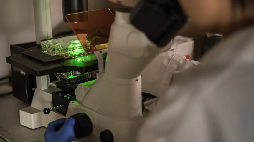
By attaching the gene encoding GFP to genes of interest, scientists can make otherwise invisible proteins visible and track them in living cells under a microscope. The method has been regarded as a breakthrough in cell biology, enabling real-time observation of molecular processes.
However, new research led by Beata Wielgus-Kutrowska, PhD, at the University of Warsaw’s Institute of Experimental Physics, challenges assumptions about the reliability of this marker. The findings were published in the International Journal of Biological Macromolecules.
Researchers have long assumed that attaching GFP to another protein does not alter that protein’s properties and that fluorescence indicates correct folding and normal behaviour.
“We also assume that when this complex glows, the GFP protein is properly folded and behaves naturally,” Wielgus-Kutrowska said in an interview with PAP.
Her team examined whether this assumption always holds true.
“Most scientists focus on what GFP illuminates in the cell, and much less often consider what happens to GFP itself. We wanted to check whether we always see what we should see. For example, whether the signal is distorted by protein misfolding or its tendency to aggregate into larger clusters, which could interfere with the reading,” she said.
Where does the green light come from?
For GFP to emit light, it must first fold into a characteristic barrel-like structure. Inside this structure, a chromophore forms — the molecular fragment responsible for fluorescence. The formation of the chromophore requires several chemical reactions within the folded protein, including ring closure, reaction with oxygen and the loss of a water molecule. Even after folding, the chromophore needs time to mature before fluorescence begins.
The researchers found that the folding process differs depending on whether GFP is folding for the first time without a chromophore or refolding after one has formed.
“It turned out that depending on whether the observed marker has already folded and has already formed a chromophore, or whether the protein is folding for the first time (without a chromophore), the process runs differently. A molecule undergoing primary folding is more prone to forming incorrect forms that stick together, but this process is less dynamic than in the case of a protein with a chromophore. However, molecules that have achieved the correct structure begin to glow later compared to those that refold,” Wielgus-Kutrowska said.
She stressed that the absence of fluorescence does not necessarily mean the absence of the labelled protein.
“The absence of glow does not always equate to the absence of a marker and may mask some rapid processes occurring in living cells,” she said.
When a protein does not fold correctly
The team also observed that GFP does not always fold correctly. In some cases, molecules aggregate into clusters that do not fluoresce. In such situations, researchers may incorrectly conclude that a protein is not present in a given location, when in fact the GFP tag is misfolded and inactive.
The findings may be relevant in studies of neurodegenerative diseases such as Alzheimer’s, where GFP is used to label proteins forming deposits in the brain. If GFP aggregates and fails to emit light, these deposits may remain undetected under a microscope.
Researchers also noted that cells may recognise GFP aggregates as abnormal and activate stress responses to remove them, potentially altering the biological processes under observation.
Molecular lightbulb
Wielgus-Kutrowska’s team concluded that both primary and secondary folding of GFP involve transitional forms with varying tendencies to aggregate and differing timelines for fluorescence appearance. This variability may distort experimental results.
Not just green
Scientists now use a broad range of fluorescent proteins in different colours. Although they differ in emission spectra, they share a similar three-dimensional structure, suggesting the problem may extend beyond classic GFP.
The researchers emphasised that fluorescent proteins remain powerful tools, but their signals should be interpreted cautiously, as the presence or absence of fluorescence may not fully reflect what is happening inside the cell.
Katarzyna Czechowicz
kap/ zan/
tr. RL